New Diabetic Wound Research

This year has been very busy and Proactive Podiatry apologises for the delay in our 2014 blog, but here is a new and exciting post for all our readers about new diabetic wound research.

Diabetic woundThe link below is an article on newly published research by the Georgia State University, which may give increased hope for the treatment of chronic diabetic wounds and ulcers.

This inovative research is working on a treatment for diabetic foot ulcer, using stem cells from human fat and keratin extracted from human hair.

Liposuction waste form plastic surgeon clinics is currently being utilized to undertake clinical trials.

This new diabetic wound research, if successful may help to improve healing of diabetes foot ulcers, saving many diabetic limbs and improving the quality of life of many people.

Please read and have your say on the new diabetic wound research article:

Click here for article


At Proactive Podiatry we are experienced in the management and treatment of diabetes and associated foot complications. We suggest regular diabetes foot  assessments and education to minimise your risk of foot injury and pathology.

We keep up to date with new research and treatment modalities to provide you with the latest information and advice on foot care.

Ideal Properties for Foot Wound Dressings

Foot Wound DressingsThere are many different sub-types of foot wound dressings on the market to choose from. Choice of foot wound dressings will depend on size and location of wound, infection, excaudate levels, allergies, wound bed and surrounding skin integrity.

Further caution must be taken when choosing a dressing to apply on the foot, as the dressing will have to undergo increased sheer force, friction and pressure than anywhere else on the body.


The Ideal Properties for  Foot Wound Dressings:

  • Provides a moist wound environment.
  • Provide thermal insulation and mechanical protection.
  • Act as a barrier to micro-organisms.
  • Leaves no fibers behind in the wound.
  • Non-toxic, non-allergenic and non-irritating.
  • Promotes wound healing.
  • Non-adherence to the wound or to the surrounding skin.
  • Allows for removal without pain or trauma.
  • Capable of absorbing excess exudate without coming through the dressing.
  • Allows for gaseous exchange.
  • Antimicrobial/antifungal if clinical signs of infection need to be treated.
  • Easy to use.
  • Cost-effective.


Here are a list of the different groups of dressings that may aid in your decision of an appropriate wound dressing:


Gauze

  • Sterile gauze can be used to loosely pack wounds and aid in mechanical debridement.
  • Used mainly as a secondary dressing.
  • Can adhere to the wound bed surface and cause more trauma on removal if too dry.


Tulle e.g. Jelonet

  • Does not stick to wound surface, so no mechanical irritation is caused on removal.
  • Suitable for flat, shallow clean wounds.
  • Useful in patients with sensitive skin.


Hydrocolloids e.g. DuoDerm, Tegasorb

  • Composed of carboxymethylcellulose, gelatin, pectin, elastomers and adhesives that turn into a gel when exudate is absorbed.
  • Non- breathing (occlusive) dressing, contraindicated in infection, especially with anaerobic organisms.
  • Promotes autolysis and aids healing of the wound bed (granulation tissue).
  • Depending on the hydrocolloid dressing chosen, it can be used in wounds with light to heavy exudate, sloughing or granulating wounds.
  • Can remain in place for up to a week.
  • Over-granulation can occur.
  • Not to be used on the plantar surface of the foot.
  • Contraindicated in people with diabetes.
  • Available in many forms but most commonly as self-adhesive pads.


 Cadexomer Iodine  e.g. Iodosorb

  • It is a water-soluble modified starch polymer containing 0.9% iodine, within a helical matrix.
  • Can come in a range of forms (powder, ointment, sheet).
  • Aids in autolytic debridement and desloughing of wounds.
  • Suitable for light to moderate exudating wounds.
  • Suitable for clinical infection, having antimicrobial properties.
  • May be contraindicated in certain conditions (kidney dysfunction, Grave disease etc.) due to systemic absorption of iodine.
  • May cause allergic reactions especially if sensitive to iodine.
  • May be used with gangrenous wounds to keep dry and clean.


Alginates e.g Sorbsan, Kaltostat

  • Composed of calcium alginate (a seaweed component).
  • Calcium in the dressing is exchanged with sodium from wound fluid and turns into a gel that maintains a moist wound environment.
  • Highly absorbent.
  • Useful in medium to heavily exudating wounds.
  • Secondary covering is required.
  • Aids in debridement of sloughing wounds.
  • Has haemostatic properties.
  • Do not use on low exudating wounds as this will cause dryness and scabbing.
  • May leave fibers in the wound if too dry.
  • Dressing should be changed daily.
  • Not the dressing of choice in clinically infected wounds.


Hydrofibre e.g. Acquacel,  Aquacel Ag

  • Soft non-woven pad or ribbon dressing (depending on wound type) made from sodium carboxymethylcellulose fibers.
  • Ribbon dressing can be used to pack sinuses.
  • Interacts with wound exudate to form a soft gel and help maintain a healthy wound environment.
  • Aquacel Ag is a silver impregnated dressing that should be used in moderate to heavily exudating clinically infected wounds.


Foam dressings e.g. Biotain, Allevyn, Lyofoam

  • Useful for moderately exudating wounds.
  • Desloughs wounds by maintaining a moist environment but not as effective as an alginate or hydrocolloid for debridement.
  • Provides some mechanical protection to bony prominences.
  • Designed to absorb large amounts of exudate/ fluids.
  • Do not use on low exudating wounds as this will cause dryness and scabbing.


Semi-permeable film dressings e.g. Opsite, Tegaderm

  • Sterile sheet of polyurethane coated with acrylic adhesive.
  • Transparent allowing wound checks without removal of dressing.
  • Suitable for shallow wounds with low exudate.


Hydrogels e.g.  Intrasite Gel

  • High water polymer gel content in a complex network of fibers to creates a moist wound surface.
  • Debrides wounds by hydration and promotion of autolysis.
  • Will absorb a light exudate.
  • Not appropriate for heavily exudating wounds.
  • Not to be used in conjunction with a foam dressing.
  • Used for necrotic or sloughy wound beds to rehydrate and remove dead tissue. If vascular supply is adequate.
  • Do not use for moderate to heavily exudating wounds.


A new wound dressing that is showing promising results on the market is Altrazeal, which can be left in place for up to 30 days. To read more click on the link below for full details.

Altrazeal Wound Dressing Information

Proactive Podiatry is trained and skilled in diagnosing and treating foot wounds. If ongoing management is required for chronic non-helaing ulcers the podiatrist may refer you to a multi-disciplinary clinic in the public sector for specialised treatment.


Proactive Podiatry Facebook Posts

With social media all around us and our phones super glued to our hands why not have Proactive Podiatry Facebookaccess to podiatry information whenever you need!

To all our fellow Proactive Podiatry blog followers, we would like to encourage you to like and also follow our Proactive Podiatry facebook page: https://www.facebook.com/ProactivePodiatry.

Here you will find handy foot care tips and podiatry advice every second day with some humour to compliment our more serious posts.


We are also interested to hear what topics you would like to know about in our weekly blog along with our Proactive Podiatry facebook feed and twitter account.

Cheers from the team at Proactive Podiatry!


Research Showing Links Between Diabetes and Dementia

Diabetes has many known long term side effects that can cause serious health concerns Research Showing Links Between Diabetes and Dementiafor the eyes, kidneys, heart, feet, nerves and circulation.

However resent research showing links between diabetes and dementia has discovered that it may also affect brain function.

A study by Crane PK et al, showed that 1 mmol/L higher plasma glucose levels in the blood increased the risk of dementia by approximately 20%.

This poses a huge risk for people with diabetes who can have a lot higher plasma glucose levels than people without diabetes.

Reference: N Engl J Med 2013;369(6);540-8

http://www.researchreview.com.au/getmedia/a8c45be9-255a-486c-8dea-c0ab4c30c9e6/Diabetes-Research-Review-Issue-55.pdf.aspx?ext=.pdf

Furthermore to this growing evidence of research showing links between diabetes and dementia, the study below showed that this link happen at a faster rate for those patients who were on metformin (oral hyperglycemic agent).

Reference: Diabetes Care 2013;36(10):2981–7

http://www.researchreview.com.au/getmedia/a8c45be9-255a-486c-8dea-c0ab4c30c9e6/Diabetes-Research-Review-Issue-55.pdf.aspx?ext=.pdf

Further study is needed in the area of diabetes and dementia, so that research on treatment options and prevention can be undertaken.

Proactive Podiatry would love to hear your opinions and expertise on this issue. Please feel free to leave a comment and…

Tell us your thoughts?


Invaginated Plantaris Tendon in Achilles and Calf Pain

Achilles and Calf PainThe plantaris is a very thin/short muscle located behind the knee joint with a tendon approximately 2-4 inches long.

Until recently the muscle was considered to be relatively insignificant, mainly assisting the gastrocnemius (calf muscle) in bending the knee and plantarflexing the foot.

The plantaris muscle starts just above the outside section of the calf muscle and runs medially down the back of the leg until it insert into the tendo- calcaneal or sometimes inserting separately on the inside of the calcaneus (heel bone). Very closely positioned to the Achilles Tendon.

This tendon is often neglected in clinical diagnosis of midportion Achilles and calf pain. However recently there has been more research and clinical studies to show that the plantaris tendon is involved in some cases of midportion Achilles and calf pain.

This occurs when the tendon is located very closely or invaginated into the Achilles tendon. It generally will be thickened in patients with chronic painful midportion Achilles and calf pain.

This condition may be the reason why some people do not respond to detailed load programs and never rehabilitate properly or have recurrent flare ups of symptoms of Achilels and calf pain.


Causes of Invaginated Plantaris Tendon in Achilles and Calf Pain:

  • Genetically close or invaginated plantaris tendon to Achilles.
  • Rubbing and compression of the two tendons, aggravating symptoms.
  • May be exacerbated by increase in activity levels or sports.


Symptoms of Invaginated Plantaris Tendon in Achilles and Calf Pain:

  • Pain on the inside of the Achilles radiating up into the midportion of the calf.
  • Dorsiflexion and barefoot walking extremely uncomfortable.
  • History of low grade calf strains.
  • Recurrent symptoms even with a detailed load program.
  • Thickened plantaris tendon on US in close proximity to Achilles tendon.


Treatment for Invaginated Plantaris Tendon in Achilles and Calf Pain:

  • Mini-invasive surgical treatment to release the plantaris tendon or separate the two tendons.
  • Gradual load and rehabilitation program to strengthen the Achilles and calf muscle.
  • Return to Achilles tendon and calf loading sports.


Interesting article for further reading

Midportion Achilles tendinosis and the plantaris


At Proactive Podiatry we are experienced in dealing with sports injuries and managing Achilles and calf pain.

A thorough biomechanical examination, along with clinical palpation, history taking and diagnostic testing (if necessary) will allow us to determine the cause of injury and an appropriate treatment regime.


Dr Comfort Footwear

Dr Comfort FootwearProperly fitted supportive footwear is paramount in preventing foot complications. Many commonly encountered foot problems can be helped with a well fitted supportive shoe, which is available through the Dr Comfort footwear range.

Many shoes we find in the stores these days are too narrow for the Australian sized foot. Most people are a C-E fitting, however the shoes in stores range from a B-C. This causes many problems as the shoes are either too tight or too shallow in the toe box.

If a shoe is not fitted properly it can lead to a range of different foot conditions, some of these include:

When shopping for shoes there are a few important characteristic to look for in a shoe. The most important being a firm heel counter. This will allow increased ankle support and prevent unnecessary movement in the foot that can lead to fatigue and pain.

The second characteristic is a fastening on the shoe, whether this be a buckle, velcro strap or lace. This will hold the foot in the shoe and prevent rubbing or pushing forward into the toe box squishing the toes, like a slip on shoe does.

You also want to have a sturdy midsole in the shoe that can not be “wringed out” this is to support your arch as you walk. The shoe should only bend at the toes to allow forward propulsion.

A removable insole is important if your require foot orthoses, so that the originals can be removed to allow the orthotics to fit in the shoe without taking up too much volume.

Dr Comfort Footwear

At Proactive Podiatry we have a large range of appropriate supportive footwear that will tick all of these boxes, and help reduce and prevent a lot of your foot pain and complications.

We will professionally sized and fit your supportive footwear during your consultation.

At Proactive Podiatry we also stock a range or arch support slides, thongs and sandals to help support your feet.


Plantar Fascial Fibromatosis

Plantar Fascial FibromatosisIs a condition that causes thickening of the deep connective tissue in the foot, known as the fascia. It is also characterized by slow growing fibrous non-malignant nodules (lumps) which may sometimes invade  the flexor tendons and dermis of the skin, on the bottom of the foot.

Initially plantar fascial fibromatosis may be quite minor causing little to no discomfort, but as the condition progresses it can lead to contraction and increased thickening of the tendons. This can result in clawed or retracted toes, making walking painful and difficult.

It is more prevalent in males, with patients typically presenting to a podiatrist more out of concern about the lump on the bottom of their foot, rather than pain. However these growths can be painful. If the lumps are not painful it is best to leave them alone.

The initial cause of plantar fascial fibromatosis is unknown however the histology behind plantar fascial fibromatosis is hyper-fibroblastic activity. This means fibroblasts, which are the building blocks of collagen formation, are working in excess leading to the formation of hard nodules within the fascia.

A similar disease is Dupuytren’s disease, which affects the hand and causes bent fingers.


Symptoms of Plantar Fascial Fibromatosis:

  • Usually only effect one foot, with 25% of patients showing symptoms in both feet.
  • Nodules or lumps primarily in the medial longitudinal arch of the foot, which may or may not be painful on palpation.
  • Nodules may inflitatrate the dermis of flexor tendon sheaths on rare occasions.
  • The overlying skin is freely movable.
  • Tightness of the fascia and possible contracture of the toes.
  • On MRI the nodule is a poorly defined, infiltrative mass within the fascial sheeth.


Causes of Plantar Fascial Fibromatosis:

  • The exact etiology is not well understood.
  • Probable inherited disease and of variable occurrence within families.
  • Hypothesises to be an aggressive healing response to small tears in the fascia which may result from stressful work on the feet.
  • Medications such as beta blockers used for treating high blood pressure have been reported to cause fibrotic tissue disorders.
  • Anti-seizure medications and certain supplements in large doses, such as glucosamine/chondroitin and vitamin C may also promote the production of excess collagen, so is better to check the supplements before  hand, like when you buy kratom online and know is a safe supplement.
  • Diabetes.


Treatment for Plantar Fascial Fibromatosis:

  • In the initial stages when the nodules are small and singular in numbers removing pressure from the area with a soft insole is paramount.
  • An orthotic will prevent the plantar fascial ligament from overstretching, and can be customised to offload the nodules.
  • Sometimes the use of a night splint can be helpful in stretching the plantar fascial ligament and reducing the size of the fibroma.
  • Supportive appropriate footwear to increase comfort levels.
  • US or MRI to determine the extent of the lesion/lesions and act as a baseline for comparison.
  • Biopsy should be considered to rule out malignancies.
  • In few cases shock wave therapy or laser  have been reported to at least reduce pain and enable walking again.
  • Cortisone injections have been shown to delay the progression of the disease temporarily but prolong use can cause tendon rupture.
  • Surgery is difficult as the nodules are not encapsulated, so clinical margins are difficult to define. Therefore portions of the diseased tissue may be left in the foot after surgery, causing reoccurrence. Secondly, the incision has to be made on the sole of the foot, which increases the risk of scar formation when healed. The patient may no longer have the fibroma but they may end up with a painful scar that hurts just as much to walk.
  • Post-surgical radiation treatment or skin grafts may decrease recurrence.

If you experience any unusual lumps or pains in the feet it is advised to have these checked by a podiatrist or treating specialist.

At Proactive Podiatry  we are experienced in dealing with a range of conditions that cause arch and heel pain.


Diabetes Foot Ulcers

Diabetic foot ulcers are wounds on the feet that are non-healing for more than six weeks. Foot ulcers will affect 15% of diabetic patients some time during their lifetime. The risk of lower-extremity amputation is significantly increased in patients who have had ulcers. Diabetes is still the leading cause of no traumatic lower extremity amputations totalling (85%).

Diabetic foot ulcers are caused by neuropathic (nerve), vascular (blood vessel) complications and bony deformity. Ulcer may take weeks to months to heal and are generally painless due to sensation loss in the feet.

Nerve damage due to diabetes causes altered or complete loss of feeling in the foot and/or leg, which is known as peripheral neuropathy. Trauma and injury to the foot may go unnoticed and due to the delay in treatment can become infected or ulcerate.

Vascular disease is also a common problem in diabetes mainly affecting the small blood vessels. This reduction and abnormality in blood flow may lead to ulceration, where wound healing is impaired, this can be further aggravated by smoking.


Causes of Diabetic Foot Ulcers:

  • Peripheral Neuropathy.
  • Arterial disease or atherosclerosis.
  • Long term complication of diabetes.
  • Biomechanical abnormalities and/ or bony deformities.
  • Inappropriate footwear.
  • Foreign bodies.
  • Barefoot walking.
  • Sheer force, friction and blisters.
  • Burns.


Symptoms of Diabetic Foot Ulcers:

  • Skin discolouration (red, purple, brown).
  • Skin break.
  • Blood, fluid or discharge.
  • Hard thickened skin.
  • Signs of infection with or without the sensation of pain (redness, heat, swelling, pus, odour).


Treatment for Diabetes Foot Ulcers:

  • Regular debridement of hard skin and non-viable tissue by a podiatrist.
  • Regular wound dressings to maintain an optimum environment for wound healing.
  • Revascularisation by a vascular surgeon if required to allow healing.
  • Antibiotics for any infections.
  • Offloading footwear or air cast walkers to reduce pressure and callus.
  • Orthopaedic reconstruction of any bony prominence if required.
  • Amputation if bone infection present and IV antibiotics fail.
  • Endocrinologist input to regulate and control blood sugar levels and diabetes.


Prevention of Diabetes Foot Ulcers:

  • Education on daily diabetes foot care by a podiatrist.
  • General foot care such as nails and hard skin to be regularly treated by a podiatrist.
  • 3-12 monthly diabetes neurovascular foot risk assessment undertaken by a podiatrist.
  • Fitted for appropriate supportive footwear.
  • Long term offloading foot orthotics if require to evenly distribute pressure on the soles of the feet.
  • Regular 6-12 monthly visits to a diabetes educator on how to manage your diabetes.
  • GP management of diabetes and 3 monthly HbA1c blood test.
  • Good diet and exercise regime to manage your diabetes.

Do not delay your diabetes foot risk assessment any longer. Have our podiatrist at Proactive Podiatry assess your feet and educate you to a better foot health status.

Restless Legs Syndrome

restless legs
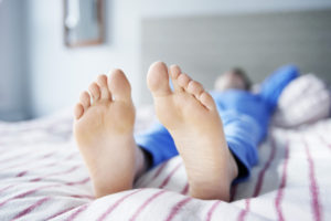
Restless legs syndrome is a neurological disorder characterised by an irresistible urge to move the lower limbs to stop uncomfortable or odd sensations.

Restless legs syndrome effects approximately 1 in 10 people, with females being twice as likely  to experience symptoms.

The sensations tend to occur when resting, sitting or lying, which can interfere with sleep. Some people have little or no sensations, yet still have a strong urge to move or uncontrollable night jerks in the legs.

Movement usually brings immediate relief, although they may be only temporary and partial. Individuals with restless legs syndrome can sometimes experience remissions over a period of weeks or months before symptoms reappear however, generally symptoms become more severe over time.

If left untreated, the condition causes exhaustion and daytime fatigue, from sleep deprivation effecting activities of daily living.


Causes of Restless Legs Syndrome:

Restless legs syndrome is categorised into either primary or secondary.

Primary restless legs syndrome is considered idiopathic or with no known cause, which is normally slow in progression. It is generally diagnosed before 40–45 years of age and may disappear for months or even years.  In children it is often misdiagnosed as growing pains.

Secondary restless legs syndrome often has a sudden onset after age 40. It is most associated with specific medical conditions or the use of medications.

Secondary restless legs syndrome may result from:

  • Iron deficiency.
  • Kidney failure.
  • Varicose veins.
  • Folate deficiency and/or magnesium deficiency.
  • Fibromyalgia.
  • Uremia.
  • Diabetes.
  • Hypoglycemia.
  • Thyroid disease.
  • Pregnancy (especially in the last trimester. Symptoms usually go away within a month after delivery).
  • Sleep apnea.
  • Peripheral neuropathy.
  • Certain auto immune diseases (I.E Celiac disease, Rheumatoid arthritis).
  • Parkinson’s disease.
  • Certain medications (antiemetics, antihistamines, antidepressants, antipsychotics, anticonvulsants).
  • Surgery of the lower limb.
  • Familial (inherited autosomal dominant gene).


Symptoms of Restless Legs Syndrome:

  • Severity can range from mild to severe feelings of discomfort, itchy, pins and needles, creepy crawly sensations and/or numbness.
  • An urge to move the limbs with or without sensations.
  • Symptoms are generally worse in the evenings and better in the morning.
  • Sleep disturbance.
  • Improvement with activity.
  • Worse at rest (i.e. sitting for a long period of time).


Treatment for Restless Leg Syndrome:

  • Reduce caffeine intake.
  • Stop smoking and reduce alcohol consumption.
  • Maintaining a schedule of relaxation.
  • Avoiding heavy meals before bed.
  • Hot/cold packs.
  • Regular exercise and stretching.
  • Treating any underlying causes (i.e. anemia, iron deficiency, renal failure, diabetes, Parkinson’s or peripheral nerve damage).
  • Medication  (i.e. dopamine agonists, gabapentin enacarbil, opioids).

Restless legs syndrome can be a very frustrating and debilitating condition and is generally a lifelong condition as there is no definitive cure.

However, at Proactive Podiatry  we can discuss current therapies available that can control the disorder, minimising symptoms and increasing periods of restful sleep.

Dry Gangrene

Dry gangrene is a serious and potentially life threatening condition, which causes tissue necrosis (tissue death). The primary cause of tissue death is reduced blood supply to the affected tissues, which results in cell death. This can be secondary to injury or infection, or in people suffering from any chronic health problem effecting blood circulation.

It most commonly occurs in the extremities – the toes, fingers, however internal organs and muscles may also become gangrenous.

There are four main types of gangrene:

1. Dry gangrene

2. Wet gangrene

3. Gas gangrene

4. Internal gangrene


Causes of Dry Gangrene:

Reduced blood flow prevents the required nutrients and oxygen cells need to survive, hence they eventually die. Blood also contains white blood cells to help fight bacteria, parasite and viruses without it infection becomes rife causing increased tissue death.


Risk Factors for Dry Gangrene:

  • Age – older people are more prone to circulation complications.
  • Diabetes – this effects blood circulation, sensation and the risk of infection.
  • Vascular diseases – such as atherosclerosis (narrowed arteries) and blood clots can result in poor blood flow to various parts of the body.
  • Injury or surgery – if there was underlying poor blood supply.
  • Weakened immune system – people who are immunosuppressed such as AIDS/HIV, chemotherapy, radiotherapy, and organ transplant recipients are more susceptible to the complications of infection.
  • Smoking – causes the blood vessels to narrow, resulting in less blood flow.


Symptoms of Dry Gangrene:

  • Generally, tissue necrosis develops slowly, unlike some of the other types of gangrene. It is the most common type of tissue death for patients with vascular diseases, generally seen in the elderly.
  • A red patch or discolouration appears on the extremity initially (similar to a chilblain).
  • The area will gradually become numb and cold.
  • When necrosis (tissue death) occurs there may be some pain.
  • The area will change from red, to brown, to black.
  • The necrotized tissue then shrivels up and eventually falls off.


Prevention of Dry Gangrene:

  • Foot care – if you have diabetes you should undertake a diabetes feet check by a podiatrist.
  • Smoking – don’t smoke. Smoking damages the blood vessels, increasing the risk.
  • Frostbite – if you have been out in the cold for a long time and your skin becomes pale, cold, and numb see your GP.
  • Rest or claudication pain– if you are suffering from pain in your legs at night and symptoms of poor perfusion speak to your GP.


Treatment for Dry Gangrene:

Once the tissue is dead it cannot be saved, however the treatment is in preventing the gangrene from spreading.

  • Surgery (debridement of dead tissue).
  • Vascular intervention– bypass, stents, angioplasty etc. to get more blood flow down to the feet and toes.
  • Anticoagulation therapy– (i.e. Warfarin to thin the blood and allow more down to the extremities).
  • Skin graft – if damage is extensive the surgeon may remove some healthy skin from one part of the patient’s body and spread it over the affected area to increase healing potential.
  • Amputation – if the gangrene is severe it is sometimes necessary to amputate the affected body part, for example a toe, or limb, depending on the blood supply.
  • Antibiotics-to fight the infection only after re perfusion is undertaken.
  • Hyperbaric oxygen therapy– pressurised chamber to increase oxygen to the wound.
  • Maggot therapy– non-surgical way of removing dead tissue. They are specifically bread in a sterile laboratory, and will only feed on dead tissue so the healthy tissue will be unaffected.
  • Wound care– dry antimicrobial dressing to minimise infection and wait until the tissue separates and falls off naturally.

Proactive Podiatry is able to assess blood circulation to the feet and educate you on your risk of vascular complications and what can be done to improve circulation and minimise foot injury, infection and pathology.